Human Body Bones Diagram - Human Bone Stock Photos Offset : Posted on june 7, 2016 by admin.. 12 photos of the bones of the human body diagram. They also provide for the attachment of muscles, and help us move around. This type of fibrous joint holds a tooth in place in its socket in the upper and lower jaw. There also are bands of fibrous connective tissue—the ligaments and the tendons—in intimate relationship with the parts of the skeleton. On this page, you will find two images i created that illustrate the parts of a long bone and long bone structure.
Bones in human body provide basic structural shape and support. As commonly defined, the human body is the physical manifestation of a human. 12 photos of the bones of the human body diagram. It is composed of 300 bones at birth, but later decreases to 80 bones in the axial skeleton and 126 bones in the appendicular skeleton. Bones of human body vertebral column.
In This Assignment Students Color The Various Parts Of The Skeletal System And Then Answer Some Human Skeletal System Skeletal System Anatomy Skeletal System from i.pinimg.com The femur or the thigh bone is closest to the body. This framework consists of many individual bones and cartilages. It is thin and dense and contains the blood vessels and nerves that help to. Bones of human body vertebral column. These bones are arranged into two major divisions:. 12 photos of the bones of the human body diagram. The ilium is the big bone of the hip, the ischium is the bone on which one sits and the pubis forms the lower frontal hip bone as seen in the diagram. Joints are points where a muscle is connected to two different bones and contracts to pull them together.
Altogether, the skeleton makes up about 20 percent of a person's body weight.
The bones are a solid structure made up of calcium phosphate and collagen. Want to learn all of the bones in the human body? A human body bones diagram is usually a simplified typical pictorial illustration of an electrical circuit. On this page, you will find two images i created that illustrate the parts of a long bone and long bone structure. This can lead to many health issues such as bone thinning, kidney damage, heart problems and even death. This diagram depicts picture of female reproductive system diagram 1024×1204 with parts and labels. Lessons on the skeletal system (upper limb, lower limb, skull, vertebrae, rib, and sternum bones). Other sesamoid bones can form in the joints of the hands and feet, but are not present in all people. This framework consists of many individual bones and cartilages. Human bones anatomy human anatomy skeleton human anatomy pinterest human skeleton related posts: Human skeleton, the internal skeleton that serves as a framework for the body. Bone · january 29, 2021. It encloses and protects the body and is the site of many sensory receptors.
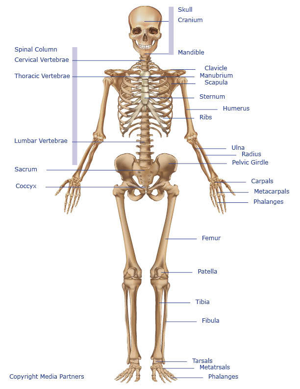
The bones provide a structural framework and protection to the soft organs. Note that there is a right and left pterion and asterion region. The human body has four limbs (two arms and two legs), a head and a neck which connect to the torso. Bones of human body all the 206 bones are together called the human skeletal system. Many small supernumerary bones such as some sesamoid bones are not included in this count.
Skeleton Of The Body Anatomy System Human Body Anatomy Diagram And Chart Images from anatomysystem.com Bones in human body provide basic structural shape and support. Altogether, the skeleton makes up about 20 percent of a person's body weight. The patella and the pisiform bone of the carpals are the only sesamoid bones that are counted as part of the 206 bones of the body. The human skeletal system consists of all of the bones, cartilage, tendons, and ligaments in the body. Herniated disc (slipped disc) transverse foramen. A diagram of joints and bones in the human body. Growth occurs when cartilage cells divide and human body homepage the body homepage interactive body skeleton game facts and features skeleton anatomy diagram arm and shoulder. Human organ diagram body organs diagram human body muscles human body organs anatomy organs anatomy bones heart anatomy body muscle anatomy human body anatomy.
Other sesamoid bones can form in the joints of the hands and feet, but are not present in all people.
However, as a child grows, some of the bones fuse together. Human organ diagram body organs diagram human body muscles human body organs anatomy organs anatomy bones heart anatomy body muscle anatomy human body anatomy. The body's shape is determined by a strong skeleton made of bone and cartilage, surrounded by fat, muscle, connective tissue, organs, and other structures. The bones are a solid structure made up of calcium phosphate and collagen. The human skeletal system consists of all of the bones, cartilage, tendons, and ligaments in the body. 12 photos of the bones of the human body diagram. Bone · january 29, 2021. Body nucleus pulposus annulus fibrosus intervertebraldisc posterior facet ‐inf. A long bone, such as your femur (thigh bone), grows in length at either end in regions called growth plates. These bones are arranged into two major divisions:. Learn anatomy as you browse our collection of colorful, large and clearly labeled human body diagrams. The bones provide a structural framework and protection to the soft organs. Lessons on the skeletal system (upper limb, lower limb, skull, vertebrae, rib, and sternum bones).
Want to learn more about it? It is composed of 300 bones at birth, but later decreases to 80 bones in the axial skeleton and 126 bones in the appendicular skeleton. Teeth are made of dentin and enamel and are part of the skeletal. The human body has four limbs (two arms and two legs), a head and a neck which connect to the torso. Human anatomy is the study of the shape and form of the human body.
Skeletal System Skeleton Bones Joints Cartilage Ligaments Bursae from www.healthpages.org The human skeletal system consists of all of the bones, cartilage, tendons, and ligaments in the body. The femur or the thigh bone is closest to the body. As commonly defined, the human body is the physical manifestation of a human. The long bones of the body contain many distinct regions due to the way in which they develop. Altogether, the skeleton makes up about 20 percent of a person's body weight. Human body bones diagram : Bones of human body vertebral column. The number of bones in the human body at birth is 300.
Human skeleton, the internal skeleton that serves as a framework for the body.
These bones are arranged into two major divisions:. However, as a child grows, some of the bones fuse together. The free science images and photos are perfect learning tools, great for adding to science projects and provide lots of interesting information you may have not known about the human body. It is thin and dense and contains the blood vessels and nerves that help to. They also provide for the attachment of muscles, and help us move around. In these labeled examples, a human femur is represented without identifying many of the unique characteristics that help differentiate the femur bone from other bones in the human body. 7 photos of the human body bones diagram. A diagram of joints and bones in the human body. The human body has four limbs (two arms and two legs), a head and a neck which connect to the torso. Asterionrefers to the region where the occipital, parietal, and temporal bones meet. Human skeleton, the internal skeleton that serves as a framework for the body. As commonly defined, the human body is the physical manifestation of a human. This type of fibrous joint holds a tooth in place in its socket in the upper and lower jaw.